Wound Healing Assay
Measures cell migration in scratch wound assays from time-lapse microscopy. Includes grid stitching for tiled image collections.
Overview
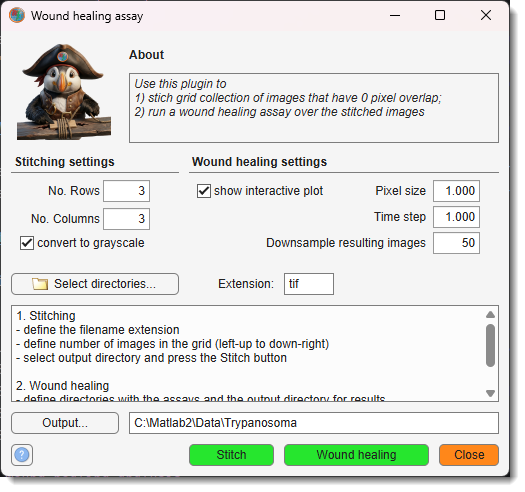
The Wound Healing Assay tool monitors cell migration into a scratch wound over time.
It operates in two independent steps:
- Stitch — assemble a tiled (grid) image collection into a single stitched image per time point
- Wound healing — detect wound width in each image and export results as plots, Excel, and MATLAB files
Launch via Ribbon → Tools → Wound Healing Assay.
Stitching settings panel
Assembles a grid of overlapping-free tile images (e.g. from Imagen Cell-IQ) into one image per time point.
- — number of tile rows in the grid (default: 3)
- — number of tile columns in the grid (default: 3)
- — average RGB channels to produce a single-channel stitched image
- — filename extension for input images (default:
tif)
Directory selection:
- — select input directories, one per tile, ordered left-to-right then top-to-bottom (i.e., row-major). The number of directories must equal No. Rows × No. Columns.
- / — type or browse to the output directory where stitched images will be saved.
— starts stitching. Each time point is saved as <original-name>_stitched.<ext>. A text file with file modification timestamps is also written to the output directory.
Tip
When the extension is jpg, a quality prompt (0–100) appears before stitching begins.
Wound healing settings panel
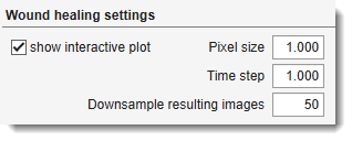
Measures wound width at each time point using the cellMigration algorithm.
- — physical pixel size in µm (default: 1). Used to convert wound widths from pixels to µm.
- — time interval in hours between consecutive images (default: 1). Used to label the time axis in result plots.
- — resizes annotated wound snapshot images to this percentage of their original size before saving (default: 50 %). Reduces disk usage for result snapshots.
- — display a live plot of wound width (min / average / max) after each time point is processed.
Directory selection:
- — select one or more directories of stitched images to analyse. Each directory is processed independently.
— runs the analysis. For each directory:
- Detects wound boundaries per image using
cellMigration.m - Plots minimum, average, and maximum wound width vs. time
- Saves annotated images to a
snapshots/subfolder inside the input directory - Exports results to
WoundAssayResults_<dirname>.matandWoundAssayResults_<dirname>.xlsx
Warning
cellMigration.m must be on the MATLAB path. It is distributed with the MIB plugin under Plugins/WoundHealingAssay/.
Exported results
| Output | Location | Content |
|---|---|---|
.mat file |
input directory | results.minVec, results.maxVec, results.avVec — wound width (µm) per time point |
.xlsx file |
input directory | Same data as a spreadsheet with image names, time points, and pixel/time step metadata |
| Snapshots | snapshots/ inside input dir |
Annotated wound images (Wound_<original-name>) |
| Timestamps | output directory | <name>_TimeStamps.txt — file modification dates for each stitched time point |
Batch scripting
Batch mode runs the Stitch step only (wound healing requires interactive cellMigration output).
Example
BatchOpt.Extension           = 'tif';
BatchOpt.NoRows              = {3, [1 Inf], 'on'};
BatchOpt.NoColumns           = {3, [1 Inf], 'on'};
BatchOpt.SelectedDirectories = {'C:\data\pos1', 'C:\data\pos2', ...};
BatchOpt.OutputDirectory     = 'C:\data\stitched';
BatchOpt.ConvertToGrayscale  = true;

obj.mibController.startController('controllers.WoundHealing', [], BatchOpt);
References
Based on Cell Migration in Scratch Wound Assays by Constantino Carlos Reyes-Aldasoro.
Cite as: CC Reyes-Aldasoro, D Biram, GM Tozer, C Kanthou, Electronics Letters 44(13), 791–793, 2008. Code available on GitHub.
Back to MIB | User interface | Ribbon | Tools