Image Converter
Back to MIB | User Interface | Plugins | File processing
The Image Converter plugin in Microscopy Image Browser (MIB) enables batch conversion of images from any MIB-supported format (e.g., NRRD, HDF5) to AM, JPG, PNG, TIF, or XML-header formats. It streamlines dataset preparation for analysis or compatibility with other tools.
Overview

The plugin provides a graphical interface to convert multiple image files efficiently.
- Support for various input and output formats.
- Optional use of Bio-Formats for advanced file reading.
- Generation of pyramidal TIFs for large datasets.
- Parallel processing to speed up conversions.
- Customizable filename prefixes and suffixes.
Usage
Access the plugin via:
Menu → Plugins → File Processing → Image Converter.
Note
Ensure input files are in a supported format. Check the supported formats list for compatibility.
Steps
-
Select Input Directory:
- Choose the folder containing images to convert .
- Enable to process nested directories.
-
Specify Input Format:
- Select the input image extension (e.g., NRRD, HDF5).
- Use for complex formats, specifying the format and series index if needed.
-
Select Output Directory:
- Choose where converted images will be saved.
-
Configure Output:
- Select the output format (e.g., TIF, PNG).
- Add optional or to output filenames.
- Enable to remove colormap data.
TIF->XML convertion
TIF->XML convertion is only implemented for Zeiss Atlas Fibics TIF files to make sure that the XML files are generated at the same location as images:
- Include subfolders is selected
- Output directory is directing to the parent folder of the one selected as the Input directory
- Add prefix or suffix
-
Pyramidal TIF Options (if TIF output):
- Check for multi-resolution output.
- Set (e.g., "1, 2, 3, 4") and type.
-
Enable Parallel Processing:
- Speed up conversion for large datasets (optional).
-
Convert:
- Click to start the process.
GUI Components
Input Panel

Input directory, specify path to source images.
, check to process subdirectories.
, input format dropdown for standard image formats
, enable Bio-Formats reader to read variety of microscopy image formats.
- a separate dropdown to specify input image format.
- series index for multi-series files.
Output Panel for standard image format

Output directory, path for converted images. In case when the structure of the input directories will be preserved in the output directory
, output format dropdown to define format for resulting images:
- JPG, Joint Photographic Experts Group format
- PNG, Portable Network Graphics format
- TIF, Tag Image File Format
- ZARR, OME-ZARR version 2 or version 3 chunked format for large datasets, see below
- XML, Extensible Markup Language suitable for metadata extraction
customize prefix text to be added before the original filename
customize suffix text to be added after the original filename
, remove colormap from output files.
, enable multi-core file conversion.
, create multi-resolution TIFs.
pyramidal downsampling factors levels for TIF output.
Scale labels to downsampling scale
The scale level in the pyramid is calculated as scaleFactor = 1/2^(levelsVec-1);
compression type for TIFs.
Output Panel for OME-Zarr
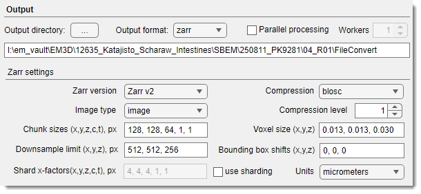
In this mode, MIB can convert files from variety formats into OME-Zarr.
Requirements and limitations
- MIB version 2.92 (beta 7) or newer
- installed Python environment with zarr-python
- the processing requires enough memory to load a sub-volume that is defined by
zChankorzChunk x zShardvalue - for parallel processing mode, make sure that you have enough memory to load as many
zChunksas many parallel processing workers defined
Loading of OME-Zarr in MIB
It is possible to open OME-Zarr version 2 or version 3 (MIB version 2.92 beta 8 or newer).
To open the dataset:
- make sure that the directory has
.zarr,.zarr2,.zarr3ending - select the directory using the
in the Directory contents panel - open the OME-Zarr dataset using the
Combine selected datasetsoption available via
, defines which version of the Zarr format to use for writing the dataset.
- 'Zarr v2' - legacy Zarr format, widely supported (e.g. MoBIE, OME-Zarr v0.4).
- 'Zarr v3' - newer Zarr format with sharding support, but fewer tools support it.
, specifies the type of data stored in the Zarr array.
- 'image' - intensity/volumetric image data (microscopy, CT, etc).
- 'labels' - segmentation or annotation data (integer label maps).
, the chunk size for storing data, written as a comma-separated list in the order [X, Y, Z, C, T]. Each chunk forms a file with these dimensions, for Zarr version 3, the chunks are merged into shards to minimize number of files. This parameter controls input/output performance: the smaller chunks give faster random access, while the larger chunks give faster sequential access.
Example
Example: '128, 128, 64, 1, 1' - chunks of 128×128×64 voxels per channel per timepoint.
, sharding factor for Zarr v3 (how many chunks are grouped together into a shard). Written as [X, Y, Z, C, T]. Reduces overhead when dealing with many small chunks, improves cloud performance.
Example
Example: '4, 4, 4, 1, 1' - each shard groups 4×4×4 chunks in XYZ per color channel and timepoint
Note: Only relevant if Zarr version = 3.
, definition of the smallest volume size until which the dataset is downsampled. For anisotropic datasets, the downsampling procedure brings the volume first to the isotropic voxels and after that downsamples all dimensions evenly.
Example
Example: '512, 512, 256' - if dataset is larger than this, downsampled levels will be generated until each axis fits within the limit.
, compression algorithm used to store chunk data.
, compression level settings: 1..9 - compression levels, the higher the value the more compression output is expected, but with the slowest computation times.
, physical voxel size of the dataset in order [X, Y, Z], given in the specified units.
Example
Example: '0.013, 0.013, 0.030' - voxel size 13 nm x 13 nm x 30 nm if units = micrometers.
, offset (translation) applied to the dataset bounding box along [X, Y, Z]. Useful when aligning multiple datasets into a common coordinate system.
Example
Example: '0, 0, 0' (no shift).
, units for physical voxel size:
Possible values
- 'nanometers'
- 'micrometers'
- 'millimeters'
- 'pixels' (unitless)
Buttons

open the Help page.
start the conversion process.
exit the plugin.
Back to MIB | User Interface | Plugins | File processing